
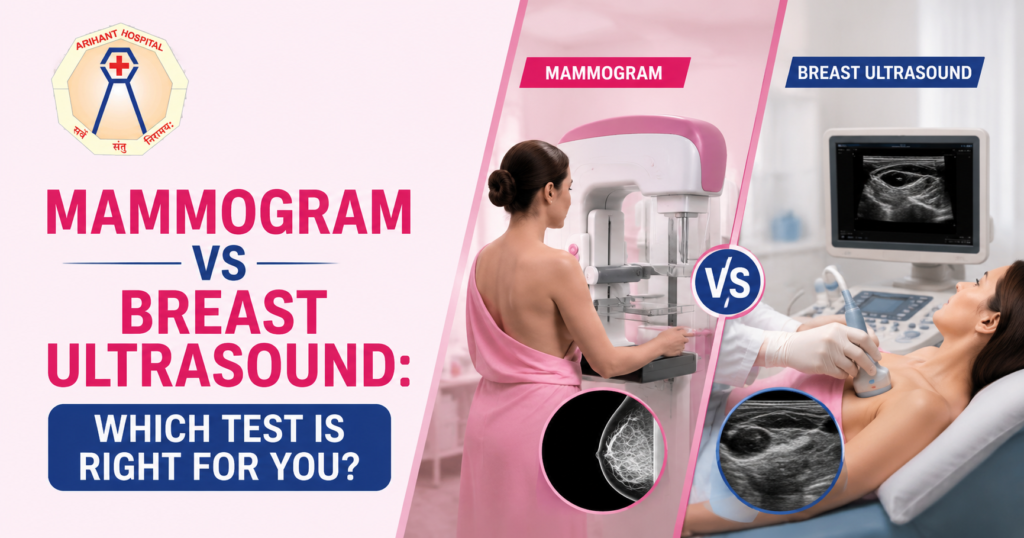
Breast health is an essential part of a woman’s overall well being, and early detection plays a vital role in identifying breast conditions before they become serious. When a lump, pain, or other breast concern arises, many women are unsure whether they need a mammogram or a breast ultrasound. While both imaging tests are valuable, they serve different purposes and are often used together to provide a more complete picture.
If you have been advised to undergo breast imaging, understanding the differences between these tests can help you make informed decisions. Consulting a qualified Breast Surgeon ensures you receive the most appropriate investigation based on your age, symptoms, medical history, and risk factors. If you are looking for Breast Cancer Treatment in Nashik, seeking expert guidance early can make a significant difference in diagnosis and treatment outcomes.
What Is a Mammogram?
A mammogram is a specialized X ray of the breast that helps detect abnormalities, including small tumors or tiny calcium deposits that may not be felt during a physical examination. It is considered the gold standard for routine breast cancer screening in women who meet the recommended age and risk criteria.
According to the American Cancer Society, mammograms can detect breast cancer before symptoms develop, allowing treatment to begin at an earlier stage when outcomes are generally better.
Mammograms are commonly recommended for:
- Women aged 40 years and above based on individual risk and physician recommendations.
- Routine breast cancer screening.
- Evaluation of suspicious breast lumps or changes.
- Follow up after previous breast abnormalities.
- Monitoring women at increased risk of breast cancer.
The procedure usually takes about 20 minutes. During the test, each breast is gently compressed between two plates to obtain clear images. Although mild discomfort may occur, it lasts only a few seconds.
What Is a Breast Ultrasound?
A breast ultrasound uses sound waves rather than radiation to create images of breast tissue. It is particularly useful for evaluating breast lumps and distinguishing between fluid filled cysts and solid masses.
Unlike mammography, ultrasound is not typically used as a primary screening tool for women at average risk. Instead, it serves as an important complementary examination.
The Radiological Society of North America explains that breast ultrasound is commonly used to evaluate abnormalities found during a mammogram or physical examination and is especially helpful in women with dense breast tissue.
Your Breast Surgeon may recommend a breast ultrasound if:
- You have a newly discovered breast lump.
- Your mammogram requires further evaluation.
- You have dense breast tissue.
- You are younger than 30 years with breast symptoms.
- You are pregnant or breastfeeding.
- A breast cyst is suspected.
Mammogram vs Breast Ultrasound: Understanding the Difference
Although both tests examine breast tissue, they are designed for different clinical situations.
| Mammogram | Breast Ultrasound |
| Uses low dose X rays | Uses sound waves |
| Best for routine breast cancer screening | Best for evaluating specific abnormalities |
| Detects microcalcifications | Excellent for distinguishing cysts from solid masses |
| Recommended for many women over 40 | Frequently used in younger women or those with dense breasts |
| May identify cancers before symptoms appear | Helps further evaluate findings from mammography |
Rather than choosing one over the other, many patients benefit from both tests depending on their symptoms and clinical findings.
Which Test Is Better for Dense Breasts?
Women with dense breast tissue often ask whether ultrasound is more accurate than mammography.
Dense breast tissue can make it harder for mammograms to identify small cancers because both dense tissue and tumors appear white on mammographic images. In these situations, your doctor may recommend supplemental imaging.
The American College of Radiology notes that additional imaging such as ultrasound or MRI may be considered in selected women with dense breasts depending on their overall risk profile.
Your Breast Surgeon will determine whether additional imaging is necessary after reviewing your mammogram results and individual risk factors.
When Might You Need Both Tests?
In many situations, mammography and ultrasound complement each other instead of replacing one another.
Common situations where both tests may be recommended:
- A lump is found during a breast examination.
- A mammogram shows an unclear area requiring further assessment.
- Dense breast tissue limits mammogram interpretation.
- Additional evaluation is needed before a biopsy.
- Monitoring previously identified breast abnormalities.
Using both imaging methods can improve diagnostic accuracy and help guide appropriate treatment decisions.
Can Breast Ultrasound Detect Cancer?
Yes. Breast ultrasound can identify suspicious masses that require further evaluation. However, ultrasound alone cannot confirm whether a lump is cancerous.
If imaging reveals suspicious findings, your Breast Surgeon may recommend a biopsy. A biopsy involves removing a small tissue sample, which is examined under a microscope to establish a definitive diagnosis.
According to the National Cancer Institute, only a biopsy can confirm whether breast cancer is present.
Are Mammograms Safe?
One of the most common concerns among patients is radiation exposure.
Modern mammography uses a very low dose of radiation, and the benefits of early breast cancer detection significantly outweigh the small risks associated with radiation exposure.
The U.S. Food and Drug Administration states that mammography remains one of the most effective tools for early breast cancer detection and uses minimal radiation.
If you are pregnant or think you may be pregnant, inform your healthcare provider before any imaging study so the safest approach can be chosen.
When Should You See a Breast Surgeon?
Many women wait until symptoms become severe before seeking medical advice. However, early evaluation is essential.
Consult a Breast Surgeon if you notice:
- A new breast lump.
- Persistent breast pain affecting one area.
- Nipple discharge, especially if bloody.
- Skin dimpling or thickening.
- Changes in breast size or shape.
- A lump under the arm.
- Persistent redness or swelling.
Not every breast lump is cancer, but every new breast change deserves professional evaluation.
Preparing for Your Breast Imaging Appointment
A few simple steps can help ensure accurate imaging results.
Before your appointment:
- Wear comfortable two piece clothing.
- Avoid deodorants, powders, or lotions on the day of your mammogram, as these may affect image quality.
- Bring previous mammogram or ultrasound reports if available.
- Inform the radiologist if you are pregnant or breastfeeding.
- Discuss any family history of breast cancer with your healthcare provider.
These small preparations help improve the quality of your examination and reduce unnecessary repeat imaging.
Early Detection Saves Lives
The choice between a mammogram and a breast ultrasound is not about which test is better but about which test is most appropriate for your individual situation. Mammograms remain the primary screening tool for many women, while ultrasounds provide valuable additional information, particularly for evaluating symptoms and dense breast tissue.
Delaying evaluation of breast changes can lead to missed opportunities for early diagnosis. If you notice anything unusual, consult a healthcare professional without delay.
Expert Breast Care at Arihant Hospital
At Arihant Hospital, Dr. Deepa Verma Lodha provides compassionate, evidence-based care for patients with breast conditions, from routine evaluations to advanced breast cancer management. Using modern diagnostic techniques and personalized treatment plans, the hospital focuses on early detection and comprehensive care.
If you are looking for an experienced Breast Surgeon In Nashik, Arihant Hospital offers expert consultation, advanced breast imaging guidance, and complete breast health services to help you make informed decisions about your care.
Hashtags
#BreastHealth #BreastCancerAwareness #Mammogram #BreastUltrasound #BreastSurgeon #EarlyDetection #BreastCancerScreening #WomensHealth #DrDeepaVermaLodha #ArihantHospital #BreastSurgeonInNashik